


Orthodontics
Dr. Michael Stosich of Deerpath Orthodontics and Family Dental explains which X-rays orthodontic diagnosis requires, how long they’re good for, and why the ones from your general dentist usually aren’t a good fit for your orthodontic treatment.
Lake Forest, IL – You’ve had X-rays at your dentist’s office for years. So why would an orthodontist need new ones? It’s a question we hear a lot, along with how long X-rays stay usable and which kind actually matter. The short answer is that orthodontics needs a different kind of imaging than the ones used by your dentist to check for cavities.
Which X-rays do I need at the consultation?
At your consultation visit with Dr. Stosich, we take a digital panoramic X-ray using the lowest radiation dosage available for patient safety. This is included in your consultation visit so Dr. Stosich can get a general global view of your teeth and jaws.
Can I use X-rays from my regular dentist?
It depends. If you have a recent panoramic X-ray taken within the previous 3 months, we can use that instead of taking a new one.
The small X-rays are a different story. The ones dentists take for things like cavities, called PAs or bitewings, are not of diagnostic quality for orthodontics. They’re the right tool for finding decay, just not for planning how your teeth and jaws should move.
What happens once I decide to move forward?
The consultation image is only the first step. Once you agree to treatment, we take a more complete set of records.
“When the patient agrees to be in treatment, a full suite of diagnostic records are taken, which includes 3-D CBCT, a lateral cephalogram and intraoral digital light photometry scan of the teeth,” says board-certified orthodontist Dr. Stosich. Extra- and intraoral photographs are taken at the consultation as well.
A few of the above imaging terms deserve a translation into layman’s terms:
| Imaging or record | Example equipment | What it shows |
|---|---|---|
| Panoramic X-ray. | 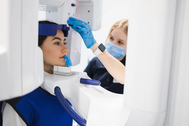 | One wide image that shows all your teeth and both jaws at once. |
| 3-D CBCT. | 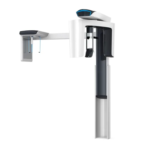 | A cone beam CT scan, which gives a three-dimensional view of your teeth and jaws. |
| Lateral cephalogram. | 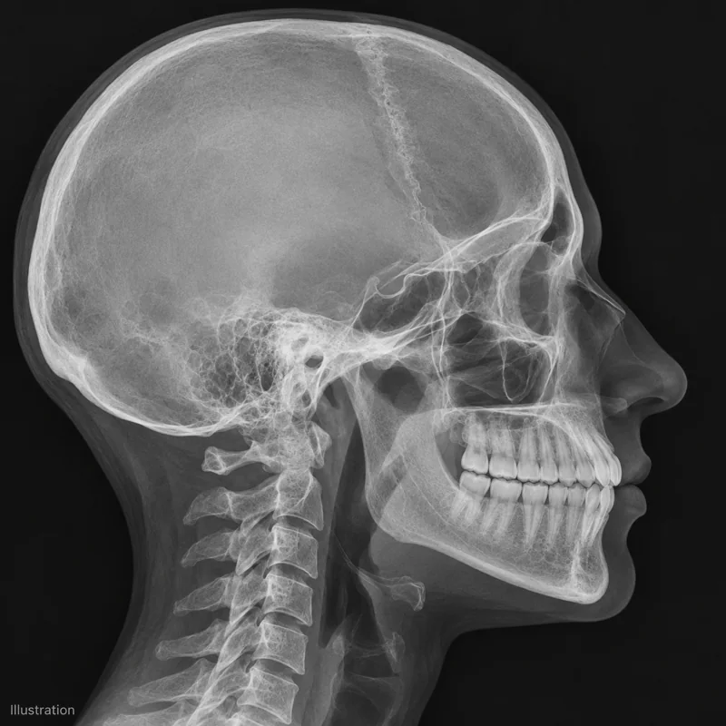 | A side-view X-ray of the head that shows how your jaws and teeth relate to each other. |
| Intraoral digital scan. | 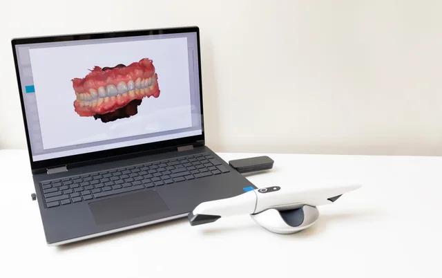 | A scan of your teeth taken inside the mouth. |
| Photographs. | 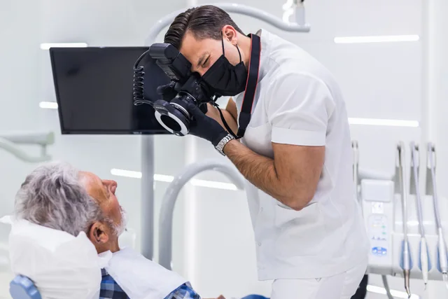 | Pictures of your face and teeth, taken at the consultation. |
How often are X-rays retaken during treatment?
Typically we do it just at the beginning and the end. In some cases, like surgical ones, a mid-treatment digital X-ray is taken too, usually a CBCT. Most patients won’t need one.
Why does it matter who reads them?
It’s because an image is only as useful as the person interpreting it. Dr. Stosich is a board-certified orthodontist (a diplomate of the American Board of Orthodontics) and the Director of Orthodontics at the University of Chicago, with more than 15 years of clinical experience. He keeps radiation low and takes only what’s needed to plan your orthodontic care properly as part of the evidence-based approach he brings to every Lake Forest patient.
If you aren’t sure whether your latest X-rays from another office will still work, bring them with you or tell us when they were taken and what kind they are, and we’ll tell you.
Call Deerpath Orthodontics and Family Dental to schedule your consultation with Dr. Stosich. Take the first step today.

